This page contains automatically translated content.
Internal clocks: The structure of the cells plays an important role
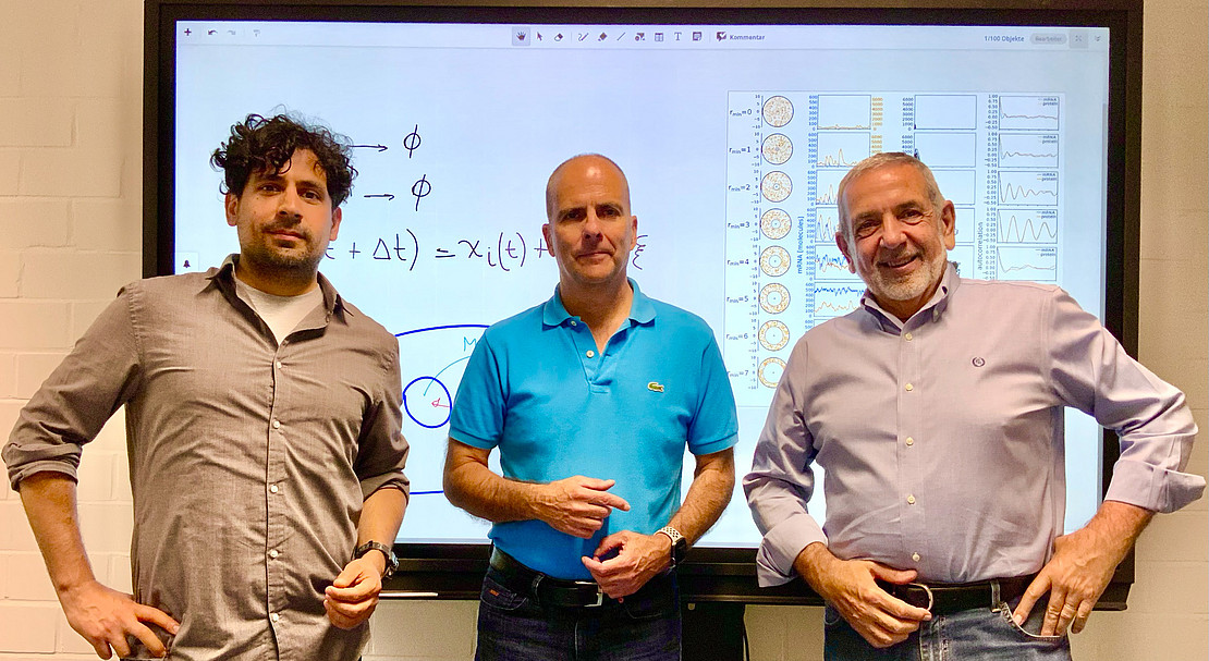 Image: University of Kassel.
The rhythms that control our daily routine depend on how our cells are organized internally, according to this fundamentally new finding. Many creatures have internal clocks that ensure they follow regular patterns, such as waking and sleeping. These clocks help them to prepare for things like foraging or mating. From tiny single-celled organisms to complex creatures, cells have evolved networks that function like chains of biochemical reactions and act as internal clocks.
Until now, modeling was based on the assumption that these clocks function like a vessel in which all the necessary chemicals are always ready to react. However, the research team, consisting of Dr. Pablo Rojas and Prof. Dr. Martin Garcia from the University of Kassel and Prof. Oreste Piro from the University of the Balearic Islands, has shown that what matters is the time it takes for a molecule to travel from its place of formation to its reaction site. The scientists developed mathematical equations that describe the individual movement of the molecules within the cell and came to the conclusion that the internal arrangement of the cell can trigger or suppress the periodic ticking of the clock.
"In the cells of eukaryotes, i.e. animals, plants and fungi, there is a physical separation between the nucleus, where the genetic code for the production of proteins is stored, and the cytoplasm, where the proteins are produced by ribosomes," explains Prof. Garcia. Although the clock cells are tiny, usually in the micrometer range, their crowded internal organization presents a path full of obstacles for the migration of molecules: The cytoplasm, for example, may itself be subdivided by membranes, and it also contains numerous elements ranging from mitochondria to lyosomes. This means that the proteins responsible for the internal rhythm take a considerable amount of time to reach the cell nucleus, where they set the clock machinery in motion. This delay is crucial for the functioning of the internal clock.
"This concept explains the existence of rhythms under conditions that were thought not to occur," says Dr. Rojas, a postdoctoral fellow in Prof. Garcia's group. "We have broken the dogma that a single expression-repression loop cannot oscillate," adds Prof. Piro, who is currently spending a sabbatical at the Institute of Physics at the University of Kassel.
From theory to application
The better and simpler models resulting from this breakthrough could eventually lead to improvements in chronomedicine, for example by developing therapies for chronopathies such as jet lag and sleep disorders, as well as optimizing the timing of drug administration in conventional treatments. But that's not all: researchers working in synthetic biology, a field that aims to build life-like circuits analogous to electronic circuits, could benefit from the development of simpler oscillating modules.
This work was carried out as part of the Research Training Group "Biological Clocks on Multiple Time Scales":
https://www.uni-kassel.de/forschung/multiscale-clocks/welcome
an interdisciplinary collaborative project funded by the German Research Foundation, in which researchers from biology, physics, mathematics, engineering and chemistry at the University of Kassel work together to research and decipher biological rhythms.
Link to the paper: https://link.aps.org/doi/10.1103/PhysRevLett.132.268401
DOI: 10.1103/PhysRevLett.132.268401